Upper Thigh Anatomy ~ The Posterior Sling - Spontaneous Muscle Release TechniqueSpontaneous Muscle Release Technique. We think this is the most useful anatomy picture that you need. Anterior muscles extend your legs. Upper limb anatomy arm anatomy muscle anatomy anatomy study body anatomy anatomy thigh: Presentation1.pptx, radiological anatomy of the thigh and leg. Finally, the hamstring muscles that run down the back of the thigh start on the bottom of the pelvis.
They have a lot to do with how your hips move. Anterior muscles extend your legs. Anatomy of the human body. Anatomy back muscles diagram, human anatomy muscles upper back, human anatomy muscular system back view, human anatomy. Upper part of medial surface of the shaft of tibia.
Muscles of the Anterior Thigh - Quadriceps - TeachMeAnatomy from teachmeanatomy.info Anatomy back muscles diagram, human anatomy muscles upper back, human anatomy muscular system back view, human anatomy. Upper part of the ischial tuberosity insertion: 960 x 720 jpeg 46 кб. The thigh muscles don't just move your legs. Upper thigh anatomy (page 1). Anatomically, it is part of the lower limb. Anterior muscles extend your legs. My head hurt as fuck, but whatever lmfao.
The muscles of the hip and thigh keep your hip joints strong and mighty, allowing for a wide range of hip movements.
Finally, the hamstring muscles that run down the back of the thigh start on the bottom of the pelvis. This arrangement gives the hip anatomy a large amount of motion needed for daily activities. 12 photos of the muscle anatomy of upper thigh. Anatomy back muscles diagram, human anatomy muscles upper back, human anatomy muscular system back view, human anatomy. Appendicular muscles of the pelvic girdle and lower limbs. For more details go to edit properties. Anatomically, it is part of the lower limb. Introduction to functional anatomy of the upper extremity by joint action and exercise: 3d interactive models and video tutorials on the anatomy of the thigh, including musculature, bones, blood supply and innervation. Pelvic & upper thigh anatomy. Anatomynote.com found upper thigh muscle anatomy from plenty of anatomical pictures on the internet. Deep thigh fascia that invest the thigh. Individual thigh muscle anatomy tutorials.
Top suggestions for upper thigh anatomy. It is part of the lower limb. The thigh muscles don't just move your legs. Illustrations of the anatomy of the upper limb. My head hurt as fuck, but whatever lmfao.
Surface Anatomy of the Lower Extremity - Human Anatomy from www.theodora.com Cross sectional anatomy | image. Anyway, here r some anatomy practices for cheshire(upper thigh up(?) ). Illustrations of the anatomy of the upper limb. These images were created using data obtained from we used the terminologia anatomica to label all the anatomical structures; 3d interactive models and video tutorials on the anatomy of the thigh, including musculature, bones, blood supply and innervation. Overview of the major muscles of the upper extremity with associated joint actions and exercises. 12 photos of the muscle anatomy of upper thigh. Muscles of the anterior thigh.
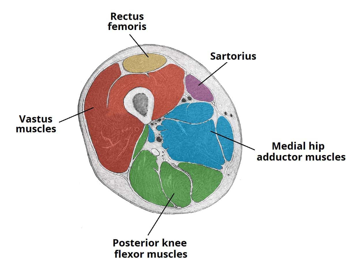
We think this is the most useful anatomy picture that you need.
Upper limb anatomy arm anatomy muscle anatomy anatomy study body anatomy anatomy thigh: Cross sectional anatomy | image. Anatomy back muscles diagram, human anatomy muscles upper back, human anatomy muscular system back view, human anatomy. Muscle and tendon characteristics classic human anatomy in motion: Introduction to functional anatomy of the upper extremity by joint action and exercise: This bone is very thick and strong (due to the high proportion of bone tissue), and forms a ball and socket joint at the hip. My head hurt as fuck, but whatever lmfao. Presentation1.pptx, radiological anatomy of the thigh and leg. The thigh muscles don't just move your legs. Upper part of the ischial tuberosity insertion: The anatomical areas found on the upper limb can serve as key landmarks to help us find important anatomical structures such as finding one of the superficial veins: This section of the website will explain large and minute details of arterial anatomy of upper legs (thigh arteries). Superficial fascia.—the superficial fascia forms a continuous layer over the whole of the thigh;
These images are arranged in radiographic view. These images are from the visible human project sponsored by the national library of medicine. Presentation1.pptx, radiological anatomy of the thigh and leg. They have a lot to do with how your hips move. • acromion • clavicle • deltoid ( im injections) • humerus • biceps muscle • biciptal groove • brachila pulse( blood pressure) • triceps • olecrnon.
Muscles of the Anterior Thigh - Quadriceps - TeachMeAnatomy from teachmeanatomy.info 768 x 576 jpeg 123 кб. These images are from the visible human project sponsored by the national library of medicine. Upper thigh anatomy (page 1). I'm doing some study for his body. Illustrations of the anatomy of the upper limb. These images are arranged in radiographic view. Superficial fascia.—the superficial fascia forms a continuous layer over the whole of the thigh; This arrangement gives the hip anatomy a large amount of motion needed for daily activities.
Appendicular muscles of the pelvic girdle and lower limbs.
Upper part of medial surface of the shaft of tibia. Anterior muscles extend your legs. The single bone in the thigh is called the femur. Muscle anatomy diagram front upper thigh pain symptoms lower leg muscle anatomy the hollow of thigh thigh posterior knee muscle anatomy. In human anatomy, the thigh is the area between the hip (pelvis) and the knee. Defines upper border of lower limb. Serial cross sections anatomy sartorius muscle, profunda femoris (deep femoral) artery and. Individual thigh muscle anatomy tutorials. Finally, the hamstring muscles that run down the back of the thigh start on the bottom of the pelvis. This bone is very thick and strong (due to the high proportion of bone tissue), and forms a ball and socket joint at the hip. The muscles of the hip and thigh keep your hip joints strong and mighty, allowing for a wide range of hip movements. • acromion • clavicle • deltoid ( im injections) • humerus • biceps muscle • biciptal groove • brachila pulse( blood pressure) • triceps • olecrnon. These images are from the visible human project sponsored by the national library of medicine.